


Smart Studio
Smart Studio
Lens Effect 3D
Lens Effect 3D
Optic Studio
Optic Studio
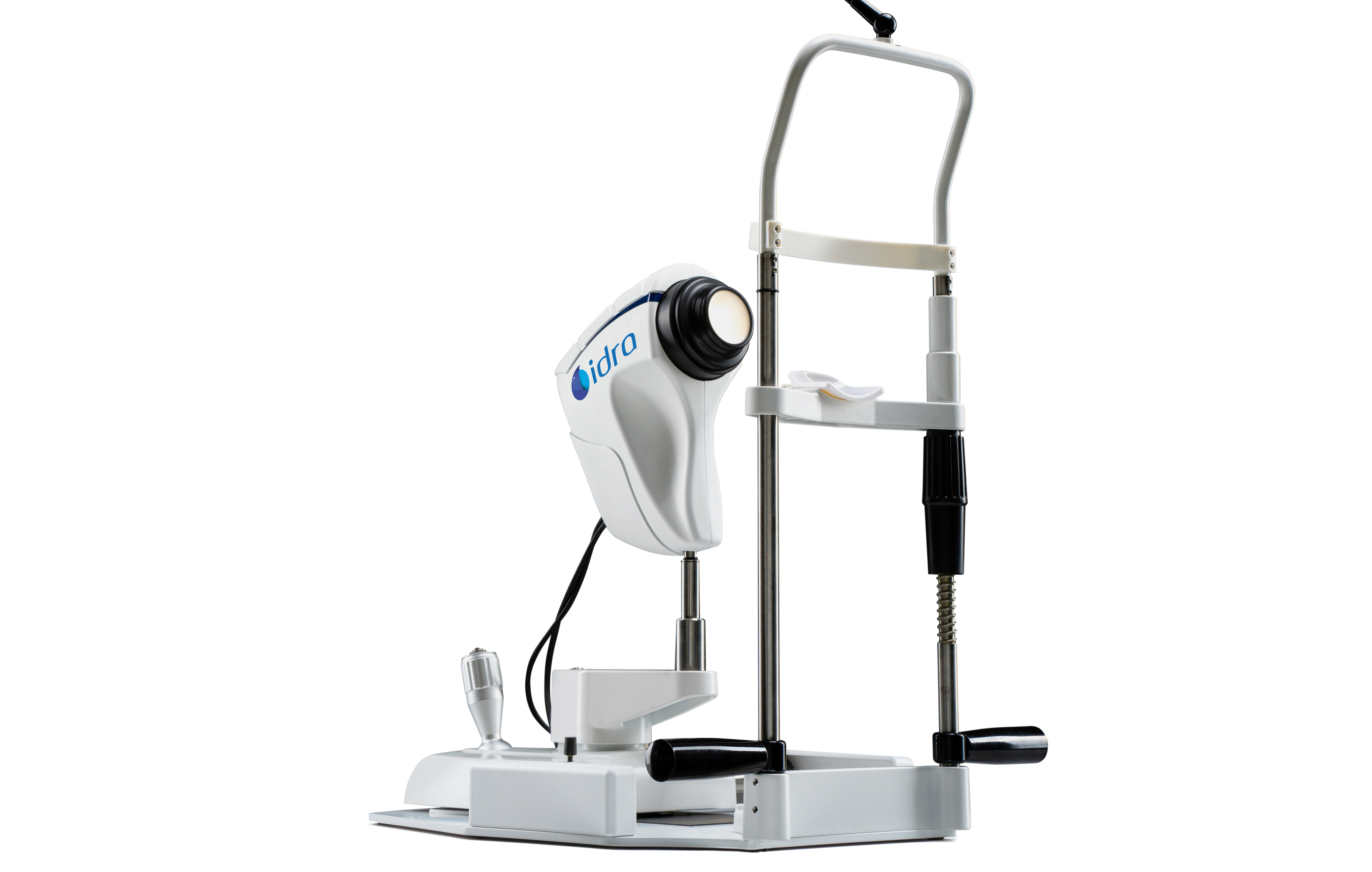
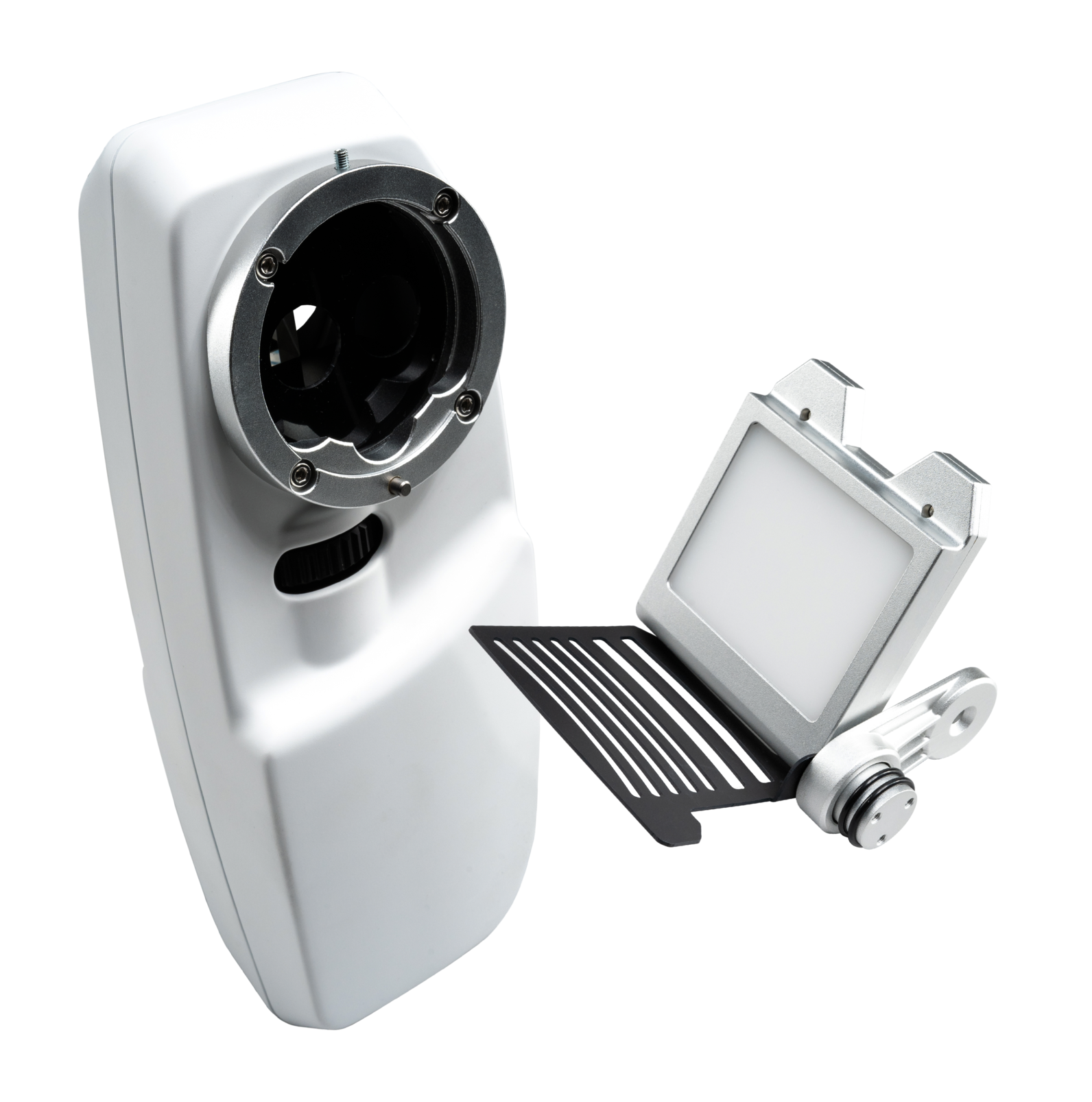
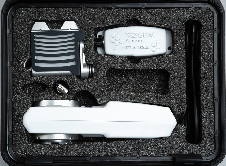
Turn your slit lamp in a complete
dry eye assessment device
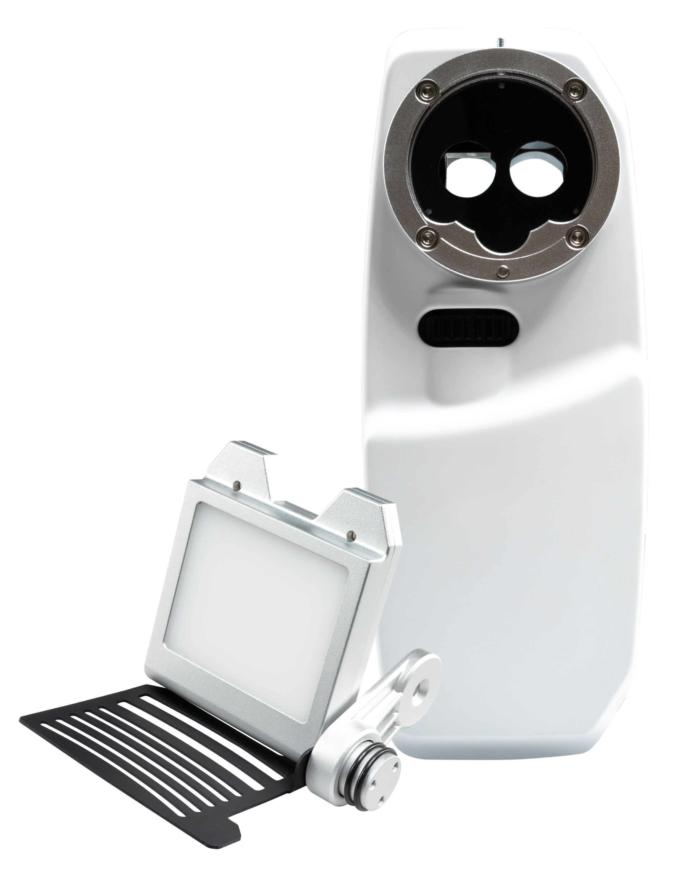
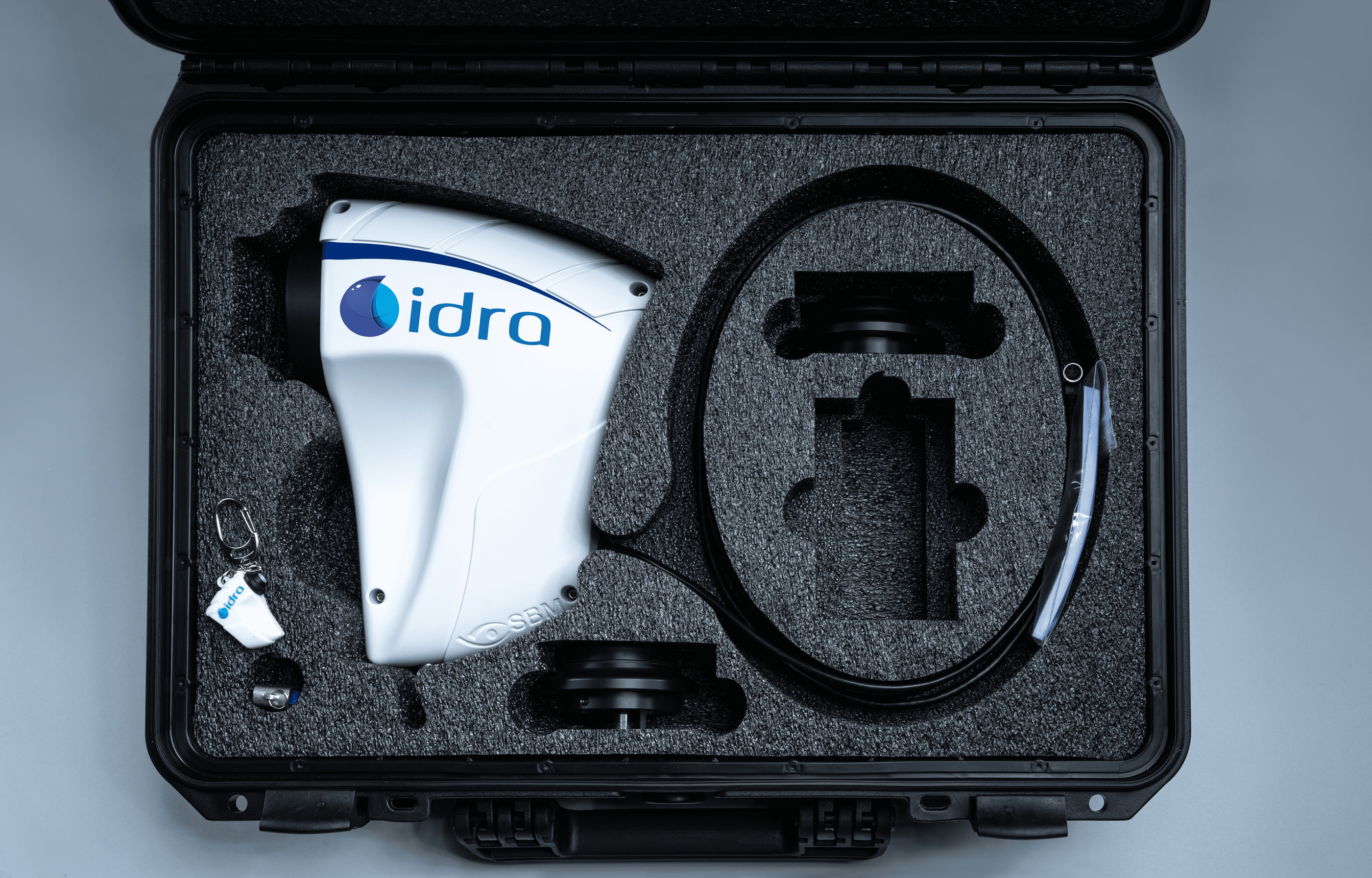
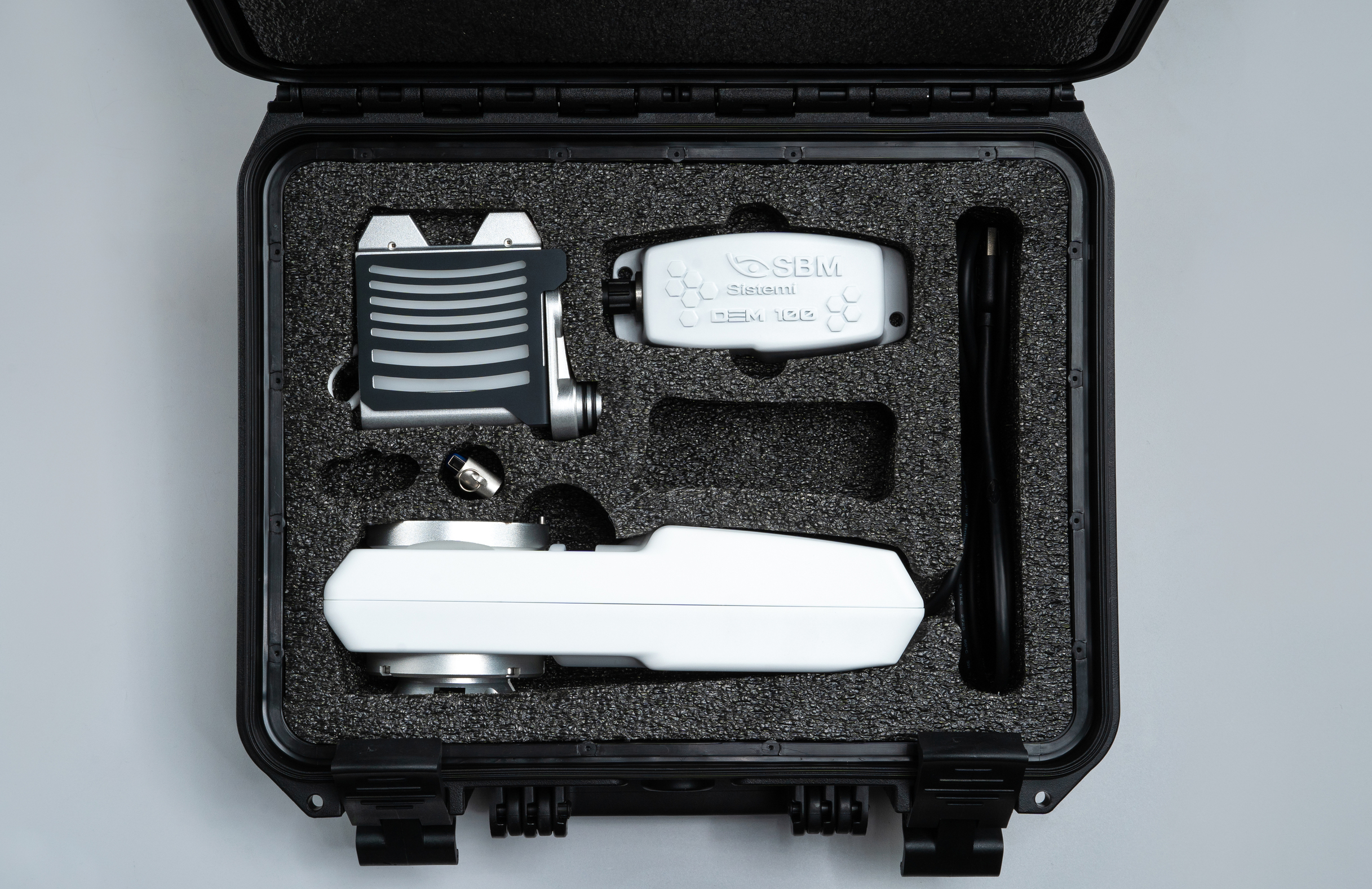
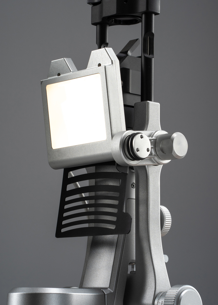
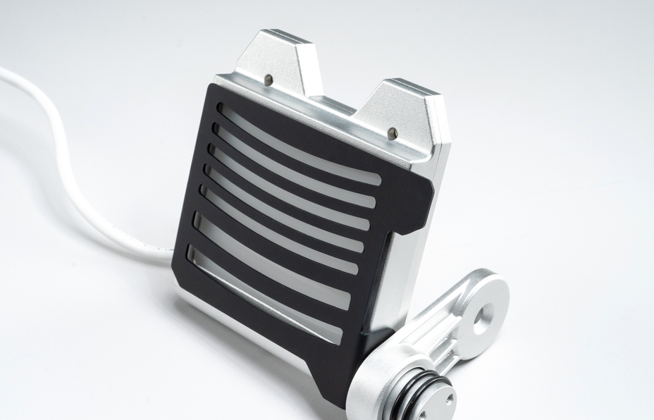
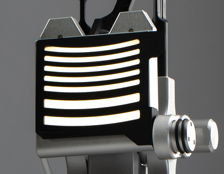
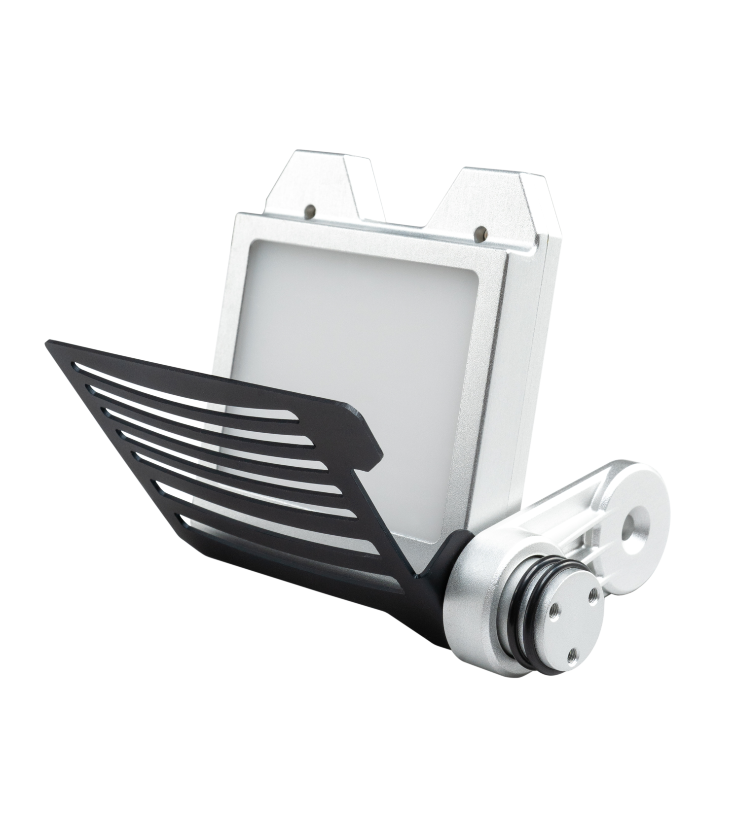
DEM100
Lighting source:
White diffuser, NIR
Lighting control:
Electronic
Power source:
USB cable
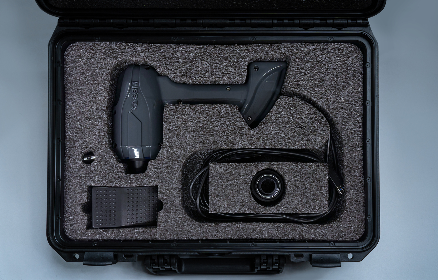
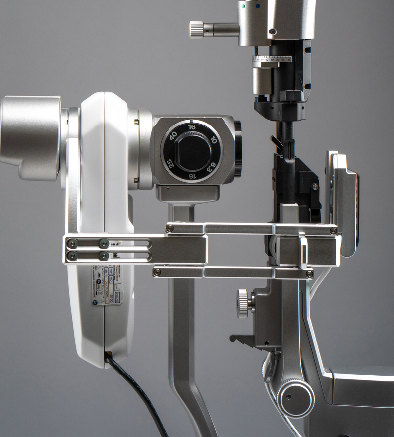
DSLC200
Acquisition card:
Not required
Image resolution:
up to 3,1 MP
1/1.8" sensor
Acquisition mode:
Single pictures,
Multi shot, Video
Focus:
Manual
ISO management:
Electronic
Connection:
USB 3.0 SuperSpeed
Many accessories,
better experience
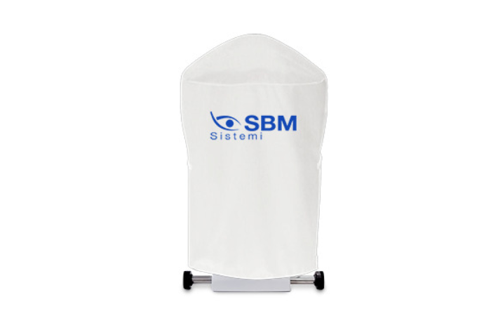
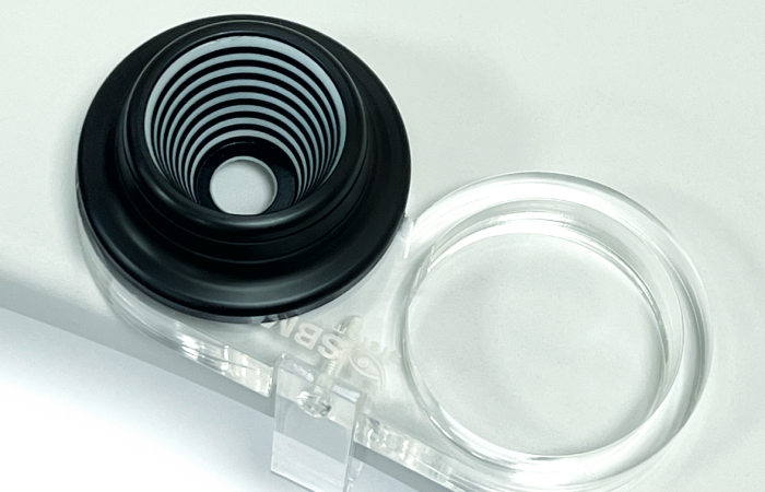
Device Cover
Cover for devices.
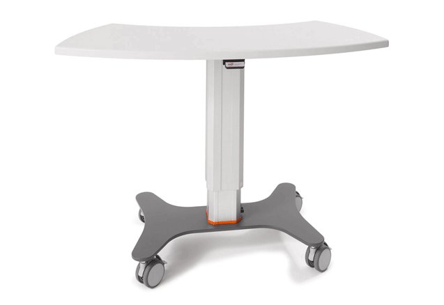
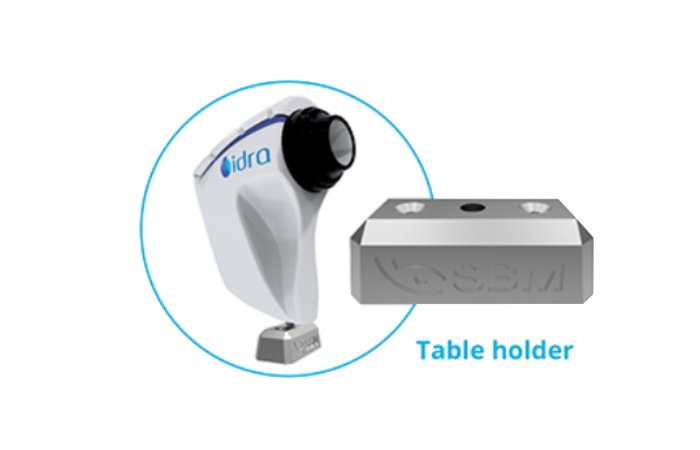
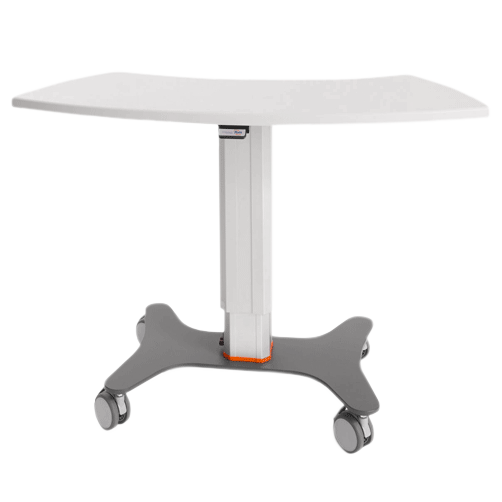
Opthalmic Table
Curved table top (V_shape) with aluminum lifting column, for 2 devices.
Table top size 1040mm x 550mm.
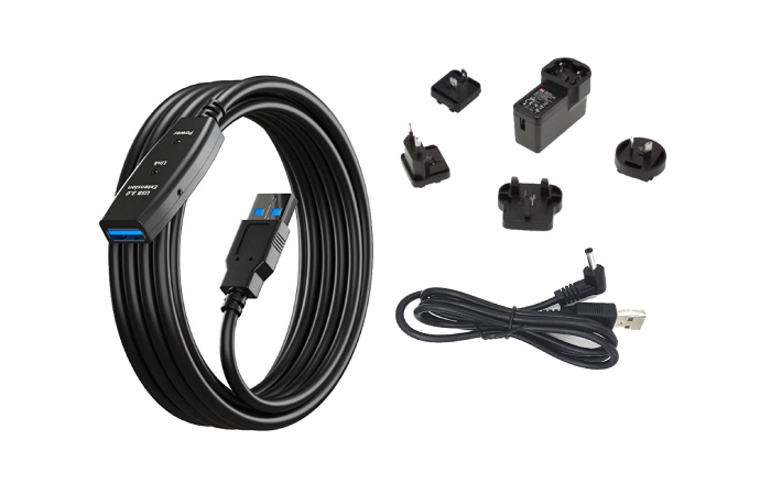
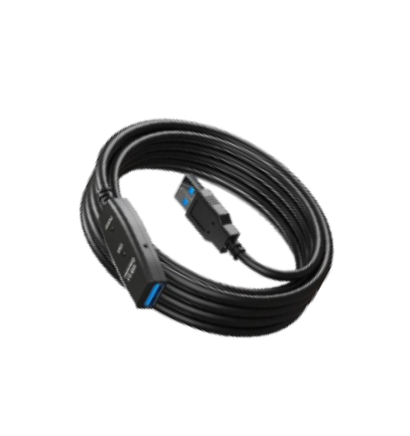
Extended USB Powered Cable
Dimensions 7 m
Powered USB 3.0 extension cable for camera to computer connection.
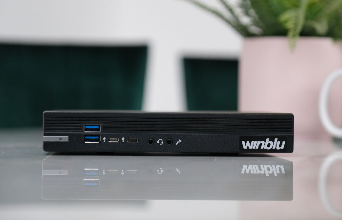
Mini Computer
Compact form factor computer conveniently configured and tested with SBM Software to properly run your new device with the convenience of a plug and play solution.
CPU: Intel® Core™ i7-12700 12 Core - 2,10/4,90 Ghz
RAM: 16GB DDR 4 (3200 MHz)
STORAGE: 512 GB SSD
GPU: Intel® UHD 770 4K - 2xDisplayPort, 1xHDMI
CONNECTIVITY: 2.5Gb LAN, Wi-Fi 6, Bluetooth 5.2
USB: 4xUSB3.2, 2xUSBtypeC, 2xUSB2.0
SOFTWARE: Windows 11 PRO
Device Cover
Opthalmic Table

Extended USB Powered Cable

Mini Computer

Opthalmic Table
Device Cover

Extended USB Powered Cable
Mini Computer
Explore numerous exam options
The revolutionary introduction of the 3D Meibomian Gland imaging gives two big advantages. Firstly, it enables to confirm the presence of abnormal glands compared to a healthy subject in a 3D view; secondly, it provides a clear image to share with the patients, to help explain the potential reason of their discomfort.
The software automatically detects the coloured lipids on the patient's eye and determines lipid layer thickness (LLT). In a few seconds it is possible to get automatically relevant data to understand functionality Of Meibomian Glands. The ICP software analyses lipid layer thickness and allows to understand the functionality of meibomian glands. lt is possible to carry out a follow up after MG treatment detecting an increase in secretion.
The human skin surface is known to house millions Of bacteria, though some people have more than the average number. Blepharitis is an inflammation caused by some bacteria that lie at the base of eyelashes. They produce dandruff-like flakes in the skin, which lead to infection and inflammation.
Acquiring an image of the conjunctiva, it will be possible to compare the patient's condition with different international grading scales.
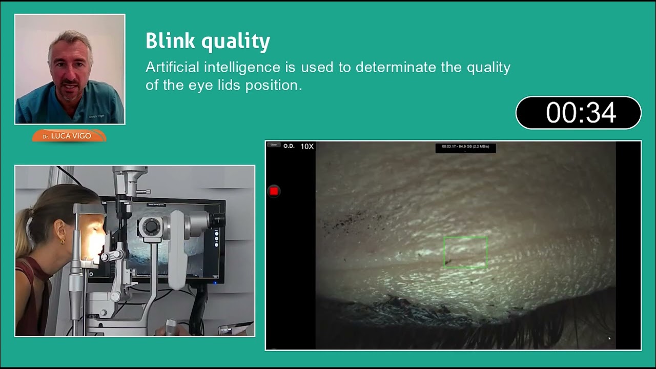
A complete blink is essential for eye health, as it refreshes the tear layer and maintains optical clarity. On average, a person blinks 11 times per minute. Incomplete blinking is often seen in contact lens users, while frequent blinking may occur to preserve the tear film's lipid layer.
The lipid layer evaluation is key in diagnosing Dry Eye, aiding in identifying its cause and informing treatment decisions. The assessment includes lipid pattern classification, thickness estimation, and incidence percentages, following Guillon & Guillon's framework. Blinking frequency, completeness, and patient history are also critical for a comprehensive evaluation.
Meibomian Gland dysfunction (MGD) is characterised by chronic, diffuse abnormalities of the Meibomian Glands and altered secretion and chemical composition of meibum. The System automatically analyses the images taken through a sensitive infrared camera (NIR) to locate the Meibomian Glands in a guided way: An exam valid both for the upper and the lower eyelids; Automatic percentage of the Meibomian Gland loss area.
Low tear production may result in aqueous tear deficiency (ATD) and cause dry eye symptoms. However, measuring the tear volume is difficult since the methods available nowadays are invasive and irritating.
The system, designed with MD Luca Vigo's expertise, enables ophthalmologists to personalize diagnostic protocols and automatically suggest treatments based on exam results. It includes a diagnosis algorithm and a treatment management feature, allowing doctors to input and prescribe various treatments. A detailed report with diagnosis and treatment recommendations can be printed directly.
Images


Discover the perfect solution for you
basic
Interferomety test
Tear Meniscus
Auto NIBUT
Meibography
-
-
Blepharitis
Ocular redness classification
Anterior segment imaging

plus
Auto Interferomety test
Tear Meniscus
Auto NIBUT
Meibography
3D Meibography
Blink quality
Blepharitis
Ocular redness classification
Anterior segment imaging