SBM Delivery

At our company, we take great care in shipping and packaging our products.
We ensure that each item is securely packed to prevent any damage during transit, and our shipping processes are designed to be both efficient and reliable.
Your satisfaction is our priority, and we strive to deliver your purchases in perfect condition every time.

SBM Privacy Policy

INFORMATION FOR DATA PROCESSING
This page describes how to manage the site in relation to the processing of personal data of users who consult it, as well as methods and purposes for processing personal data.
This is an information notice issued pursuant to art.13-14 of the EU Reg. 2016/679 to those who interact with web services accessible by telematic means starting from the address: www.sbmsistemi.com
The information is provided only for the site in question and not for other websites that may be consulted through our links, of which SBM SISTEMI SRL is in no way responsible.
SBM SISTEMI SRL acting as Data Controller of your personal data, pursuant to and for the effects of the EU Reg. 2016/679 – GDPR, hereby informs you that the aforementioned legislation provides for the protection of data subjects regarding the processing of data personal data and that this treatment will be based on principles of correctness, lawfulness, transparency and protection of your privacy and your rights.
Your personal data will be processed in accordance with the legislative provisions of the aforementioned law and the confidentiality obligations laid down therein.

TYPES OF DATA PROCESSED
Navigation Data
The computer systems and software procedures used to operate this website acquire, during their normal operation, some personal data whose transmission is implicit in the use of Internet communication protocols. This is information that is not collected to be associated with identified individuals, but which by their very nature could, through processing and association with data held by third parties, allow users to be identified. This category of data includes IP addresses or domain names of the computers used by users connecting to the site, the addresses in the Uniform Resource Identifier (URI) notation of the requested resources, the time of the request, the method used to submit the request to the server, the size of the file obtained in response, the numerical code indicating the status of the response given by the server (success, error, etc.) and other parameters relating to the operating system and the user’s computer environment. These data are used for the sole purpose of obtaining anonymous statistical information on the use of the site and to check its correct functioning and are deleted immediately after processing. The data could be used to ascertain responsibility in case of hypothetical computer crimes against the site.

Data provided voluntarily by the user
The optional, explicit and voluntary sending of e-mails to the addresses indicated on this site, as well as the completion of the forms for sending specific requests entail the subsequent acquisition of the sender’s address, necessary to respond to requests, as well as any other personal data included in the message or in the form.
Specific summary information is shown or displayed on the pages of the site prepared for particular services on request.

CONSEQUENCES OF THE REFUSAL OF DATA PROVIDE
Apart from that specified for navigation data, the user is free to provide personal data contained in the request forms, to subscribe to the mailing list or otherwise indicated in contacts to request the sending of informative, technical or commercial material or other communications. Failure to provide such data may make it impossible to obtain what has been requested.

PROCESSING METHODS
The data are mainly processed with electronic and IT tools and stored both on computer media and on hard copy and on any other type of suitable support, in compliance with the methods set out in art. 6, 32 of the GDPR and through the adoption of the appropriate security measures. to prevent the loss of data, illicit or incorrect use and unauthorized access.
We inform you that, to provide a complete service, our portal may contain links to other websites, not managed by us. We are not responsible for errors, contents, cookies, publications of illicit moral content, advertising, banners or files that do not comply with current regulations and compliance with the Privacy legislation by sites managed by us to which reference is made. To improve the service offered, an immediate notification of malfunctions, abuse or suggestions to the e-mail address is welcome: info@sbmsistemi.com
Your data will be processed only by personnel expressly authorized by the Owner.

PURPOSE OF THE TREATMENT
Data processing will be carried out for:
give the possibility to access the public and / or reserved sections of the site;
view, edit, delete data related to the account;
to execute the activation and maintenance of any services signed on-line;
carry out the obligations required by laws or regulations;
the protection of the owner in court;
send or send from information subjects explicitly in charge of informative, commercial or promotional information concerning the services of interest to them allow constant monitoring of the effectiveness of the proposed service.

SUBJECTS TO WHICH PERSONAL DATA WILL BE DISCLOSED
Personal data relating to the processing in question may also be disclosed to parties to whom the right to access your personal data is recognized by law or secondary and / or community regulations. Furthermore, some data may be communicated and disseminated to internet operators of which SBM SISTEMI SRL makes use of its own domains. The data could be communicated to specific subjects to process the sending of material and products. The dissemination of collected data is not foreseen.

DATA CONSERVATION PERIOD
Please note that, in compliance with the principles of lawfulness, purpose limitation and data minimization, pursuant to art. 5 of the GDPR, the retention period of your personal data is: for the time necessary to perform the requested services.

RIGHTS OF THE INTERESTED
Reg.to UE 2016/679: Art. 15, 16, 17, 18, 19, 20, 21, 22 – RIGHTS OF THE INTERESTED
The interested party has the right to obtain confirmation of the existence or not of personal data concerning him, even if not yet registered, and their communication in intelligible form.
The interested party has the right to obtain the indication:


The interested party has the right to obtain:


The interested party has the right to object, in whole or in part:


THE HOLDER OF THE TREATMENT
The data controller is SBM SISTEMI SRL, Strada Torino 43 – Orbassano
The user has the right to obtain from the holder the cancellation (right to be forgotten), limitation, updating, rectification, portability, opposition to the processing of personal data concerning you, and in general can exercise all the rights foreseen by the articles 15, 16, 17, 18, 19, 20, 21, 22 of the GDPR.
To exercise the rights listed above, the interested party must send a written request by e-mail. The e-mail must be addressed to: info@sbmsistemi.com

Smart Base

A complete stand alone support system.

Cover

Cover for devices.

Holder

Can be applied on the desktop with two screws to store devices when not use.

Cones Holder

Can be applied on the desk to store the cones when not in use.

Opthalmic Table

Curved table top (V_shape) with aluminum lifting column, for 2 devices.
Table top size 1040mm x 550mm.

Extended USB Powered Cable

Dimensions 7 m
Powered USB 3.0 extension cable for camera to computer connection.

Mini Pc

Compact form factor computer conveniently configured and tested with SBM Software to properly run your new device with the convenience of a plug and play solution.

CPU: Intel® Core™ i7-12700 12 Core - 2,10/4,90 Ghz
RAM: 16GB DDR 4 (3200 MHz)
STORAGE: 512 GB SSD
GPU: Intel® UHD 770 4K - 2xDisplayPort, 1xHDMI
CONNECTIVITY: 2.5Gb LAN, Wi-Fi 6, Bluetooth 5.2
USB: 4xUSB3.2, 2xUSBtypeC, 2xUSB2.0
SOFTWARE: Windows 11 PRO

Ipad Apps

Corneal Topographer

Complete evaluation of the ocular surface
and dry eye management

Rings:

24

Measured points:

8640

Camera resolution:

5 Megapixel

Photo resolution:

2592x1944
JPEG format

Upscaled analyzed
image resolution:

23 Megapixel

Acquisition mode:

Single shot,
multishot, video

Focus:

Manual focus

ISO management:

Variable

Image Color:

Colours - Infrared (IR)

Lighting source:

Infrared led - White
led - Blue led

Working distance:

60mm - 90mm from
the center of the placid

Output 1:

USB 3.0

Electromagnetic
compatibility (EMC):

IEC 60601-1-2 (2015)

Supply voltage:

24V

Device operating voltage:

24V - 5V

Dimensions:

40cm (L) x 60cm (A)
x 45cm (P)

Weight:

12 Kg

Accuracy:

Class A according to UNI EN ISO 1980-2021

Many accessories,
better experience
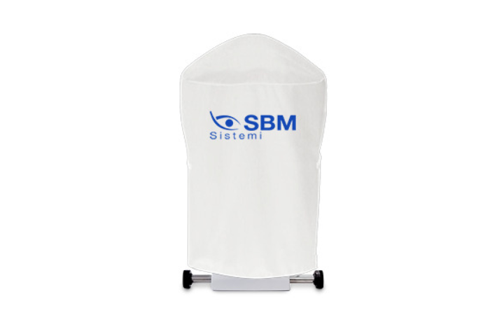
Device Cover

Cover for devices.

Opthalmic Table

Curved table top (V_shape) with aluminum lifting column, for 2 devices.
Table top size 1040mm x 550mm.

Extended USB Powered Cable

Dimensions 7 m
Powered USB 3.0 extension cable for camera to computer connection.

Mini Computer

Compact form factor computer conveniently configured and tested with SBM Software to properly run your new device with the convenience of a plug and play solution.

CPU: Intel® Core™ i7-12700 12 Core - 2,10/4,90 Ghz
RAM: 16GB DDR 4 (3200 MHz)
STORAGE: 512 GB SSD
GPU: Intel® UHD 770 4K - 2xDisplayPort, 1xHDMI
CONNECTIVITY: 2.5Gb LAN, Wi-Fi 6, Bluetooth 5.2
USB: 4xUSB3.2, 2xUSBtypeC, 2xUSB2.0
SOFTWARE: Windows 11 PRO

Device Cover

Opthalmic Table

Extended USB Powered Cable

Mini Computer

Opthalmic Table

Device Cover

Extended USB Powered Cable

Mini Computer

Explore numerous exam options

Interferomety

Tear Meniscus

3D Meibography

Meibography

Topography

Automatic N.I.B.U.T.

White to White Measurement

Bulbar Redness Classification

Pupillometry

MD Luca Vigo Suggestions

Automatic Interferomety

Automatic Tear Meniscus

3D Meibography

Meibography

Topography

Automatic N.I.B.U.T.

Blepharitis and
Cylindrical dandruff

Bulbar Redness Classification

Pupillometry

White to White Measurement

MD Luca Vigo Suggestions

full plus

3D Meibography

The revolutionary introduction of the 3D Meibomian Gland imaging gives two big advantages. Firstly, it enables to confirm the presence of abnormal glands compared to a healthy subject in a 3D view; secondly, it provides a clear image to share with the patients, to help explain the potential reason of their discomfort.

For the first time, a 3D image can help to understand the structure of the eyelids. lt can show possible diseases of Meibomian Glands and differences with healthy MGs. Patients can see for themselves why they are getting eye discomfort and fluctuating vision. Patients can understand why a specific treatment is suggested.

To be able to View the presence of abnormal gland structures in a high-resolution 3D image. Evidence that supports the diagnosis in the case of evaporative dry eye disease and the explanation of the reasons for the choice of MGD therapy (including IPL).

Automatic Interferometry

AUTOMATED LIPID LAYER ANALYSIS
The OS1000 software analyses lipid layer thickness and allows to understand the functionality of Meibomian Glands. lt is possible to carry out a follow up after MG treatment detecting an increase in secretion.

A typical blink pattern can be observed as approximately one blink every five seconds, ie 11 blinks per minute. Incomplete blink can often be observed in contact lens wearers, and frequent blink may be a result of an attempt to maintain a relatively thin lipid layer.

The software automatically detects the coloured lipids on the patient's eye and determines lipid layer thickness (LLT). In a few seconds it is possible to get automatically relevant data to understand functionality of Meibomian Glands.

Automatic Tear Meniscus

Tear meniscus prism height analisys automatically evaluated to understand main lacrimal gland functionality and detection of aqueous dry eye.

Belpharitis and Cylindrical Dandruff

The human skin surface is known to house millions of bacteria, though some people have more than the average number. Blepharitis is an inflammation caused by some bacteria that lie at the base of eyelashes. They produce dandruff-like flakes in the skin, which lead to infection and inflammation.

Problems with the Meibomian Glands (meibomianitis) in the eyelids can also cause blepharitis. The development of inflammation is also associated with tisk factors such as dandruff, dry eye, acne rosacea, or bacteria.

This test helps in the detection of blepharitis. lt can be performed on the outer surface of the eyeball and eyelids. The process includes: Examining the openings of the Meibomian Glands, base of the eyelashes, and eyelid margins using a bright light. Checking for abnormalities by evaluating the quantity and quality of tears.

The type of blepharitis can be deterrnined based on the appearance of the eyelid edges. If the symptoms frequently exhibited by the patients are mildly sticking eyelids, thickened lid margins, and missing/misdirected eyelashes, then the type of blepharitis is diagnosed as Staphylococcal.

When the patient iS found with blockage Of the Meibomian Glands in the eyelids, poor quality of tears, and redness of the lining of the eyelids, Meibomian blepharitis is diagnosed. If a hard, matted crust is formed on the eyelashes, and after its removal small sores appear on the skin, Ulcerative blepharitis is diagnosed.

WHAT IS DEMODEX BREVIS?
Demodex brevis is a kind of mite found on the skin of humans. Like its counterpart Demodex folliculorum, D. brevis is naturally occurring. D. brevis is so small that mites can't be seen macroscopically.

Interferometry

Thanks to the anterior illumination module, OSI 000 can aquire the lipid layer secrection on the cornea. The device highlights the lipid layer and the software allows to subjectively evaluate the quantity and quality of the lipid component on the tear film.

Meibography

Meibography is the visualization of the glands through illumination of the eyelid with infrared light. lt images the morphology of the glands in order to diagnose any meibomian gland drop out which would lead to tear dysfunction. Using IR illumination OSI 000 can automatically detect:
• Lid area
• Meibomian glands
• Drop out

The System automatically analyses the images taken through a sensitive infrared camera (NIR) to locate the Meibomian Glands in a guided way:
- An exam valid both for the upper and the lower eyelids
- Automatic percentage of the Meibomian Gland loss area.

Meibomian Gland dysfunction (MGD) is characterised by chronic, diffuse abnormalities of the Meibomian Glands and altered secretion and chemical composition of meibum.

The Os1000 allow an accurate comprehension of the ocular surface and especially the Meibomian Glands. The acquired images are processed and transformed into 3D pictures. Thanks to scientific algorithms it is possible for the physician to see these 3D images, and to show them and explain abnormalities to the patients.

Automatic N.I.B.U.T.

Thanks to one single video, the physician can gain lots of information:
- Automatic NIBUT
- Average of more than one value
- Graph to understand the trend of tear film stability during the video
- Tear topography that shows all breaking the tear film during time.

The stability of the mucin layer and the whole tear film is assessed through the study of non-invasive break up time (NIBUT), by using the Placido rings projected onto the cornea. Tear film stability automatically evaluated without fluorescein:
- First NIBUT
- Average NIBUT
- NIBUT Map
- TF dinamic graph

DYNAMIC PUPILLOMETRY AND WTW Evaluatlon of corneal diameter from limbus to limbus (white-to-white distante, WTW).

The measurement of the pupil diameter has become increasingly important in the field of refractive surgery. Larger scotopic pupil sizes may be partially responsible for the occurrence of postoperative symptoms such as halos, glare, and monocular diplopia.

Refractive surgeons also need an accurate scotopic pupil measurement to determine appropriate treatment zones for excimer laser, corneal, and intraocular surgery.

Bulbar Redness Classification

Acquiring an image of the conjunctiva, it will be possible to compare the patient's condition with different international grading scales.

Tear Meniscus

The thickness of the tear meniscus that is observed on the eyelid margins provides useful information on the tear volume. The tear meniscus can be examined considering its height, regularity and shape.

Topography

COMPARING EXAMS / DIFFERENCIAL MAP
Comprhensive summary veiw including elevation / tangential / saggital / refractive power maps with keratometry keratorefractive and shape indices numerical values.

SINGLE MAP
Show in detail results of specific topographic map.

3D
3D visualization of topography maps for an accurate teaching of your patient.

ADVANCED ALTIMETRY
Possibility to refer corneal shape to the best fit preffered and zernike order visualization.
Spherical - aspherical - ashperotoric surface can be selected automatically or manually set up.

ABERROMETRY
Corneal aberrometry can be evaluated on different wave front dimension to evaluate aberration according to zernike orders with the possibility to compare different corneal aberrometry.

VISUAL ACUITY
OSI 000 provides a simulation of the visual acuity trough the selected diameter of the cornea.
Visual rapresentation of PSF-3D

LENS FITTING
Virtual contact lens fitting trough manufacturer database and possibility to export topography results for a custom lens production.
Lens can be selected automatically or manually according to TLT and lens feature.

COMPARATIVE
Compare topography maps and results with previous examination.

DIFFERENTIAL
Prove the efficacy of the refractive procedure showing the changes on patient cornea.

COMPARATIVE ABERROMETRY
Compare wave front aberrometry pre and post refractive surgery or orthoK to show and prove the results to your patient.

MD Luca Vigo Suggestions and custom protocols

(MD L.VIGO Studio Carones Milano) is already provided and suggest treatment on our KOL experience.

All users can customize their own protocol adding treatment procedure to be chosen automatically right after performing the exams (this makes it possible as well to delegate the diagnosis to an assistant).

DATA RESULTS VIEW
A complete and dry eye-focused database allows to understand and properly diagnose the dry eye patient. With the useful data, the ophthalmologist can check the complete tear film assessment, determining all deficiencies and understanding which treatment is needed to approach each case.

DIAGNOSIS SUGGESTION
Ocular surface data and pathology classification. Thanks to Studio Medico Carones with MD. Luca Vigo's experience, the IDRA includes a suggestion algorithm able to share a possible treatment approach for each patient.

TREATMENT MANAGING
Through TREATMENT MANAGING tab, the software allows the physician to fill in the database with all drugs, integrators and treatments available in his practice. Any treatment as vitamins, Omega-3, eye drops, hot packs and IPL/Radiofrequency, can be loaded on the software to prescribe the products of the brands that the doctor prefers.
The whole report with the diagnosis and treatment suggested by the ophthalmologist will be printed directly.

MD. VIGO TREATMENT SUGGESTION
Suggestions for diagnosis and treatment based on clinical procedure of Dr. Luca Vigo and studio Carones (Milan, Italy)

White to White

Evaluation of corneal diameter from limbus to limbus (white-to-white distance, WTW).

Images

Videos

Discover the perfect solution for you

plus

Topography

Keratoconus screening

Contact lens fitting simulation

Pupillometry

White to white measurement

Interferometry

Auto NIBUT

Meibography

3D Meibography

Tear Meniscus

-

-

Ocular redness classification

Wizard procedure

Treatment protocol section

Smartphone App "Dry Eye Follow-Up"

OSDI

full

Topography

Keratoconus screening

Contact lens fitting simulation

Pupillometry

White to white measurement

Auto Interferometry

Auto NIBUT

Meibography

3D Meibography

Auto Tear Meniscus

Auto Blink Quality

Blepharitis

Ocular redness classification

Wizard procedure

Treatment protocol section

Smartphone App "Dry Eye Follow-Up"

OSDI

OS1000

Corneal Topographer

Request a quote

Brochures

Learn More

Report

Learn More