The Fastest and most
Comprehensive Dry Eye suite
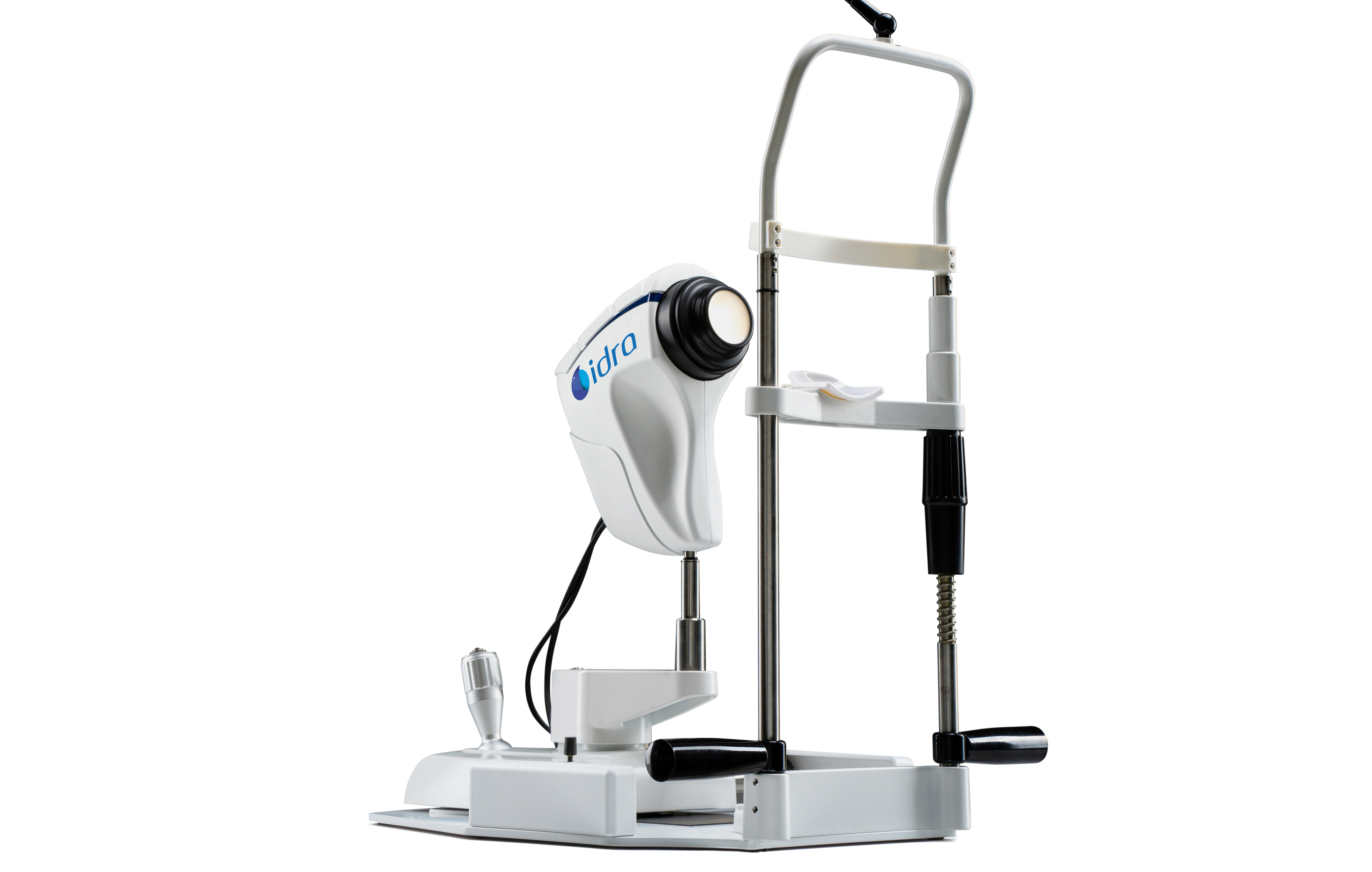
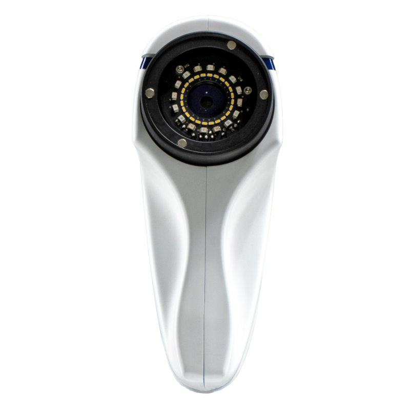
Idra is the innovative instrument
for individual tear film analysis, enabling
a rapid and detailed structural assessment
of tear composition.
Image Resolution:
8 MP
Focus:
Auto, Manual
Acquisition Mode:
Multi shot, video
ISO Management:
Variable
Camera:
Colored, sensitive
to infrared (NIR),
yellow-filtered
Cones:
Main cone,
Placid cone
Light Source:
Infrared led - blue,
red and white led

Many accessories,
better experience
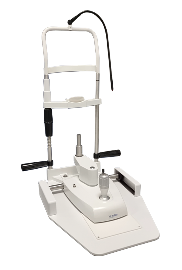
Smart Base
A complete stand alone support system.

Device Cover
Cover for devices.

Holder
Can be applied on the desktop with two screws to store devices when not use.

Cones Holder
Can be applied on the desk to store the cones when not in use.

Opthalmic Table
Curved table top (V_shape) with aluminum lifting column, for 2 devices.
Table top size 1040mm x 550mm.

Extended USB Powered Cable
Dimensions 7 m
Powered USB 3.0 extension cable for camera to computer connection.

Mini Computer
Compact form factor computer conveniently configured and tested with SBM Software to properly run your new device with the convenience of a plug and play solution.
CPU: Intel® Core™ i7-12700 12 Core - 2,10/4,90 Ghz
RAM: 16GB DDR 4 (3200 MHz)
STORAGE: 512 GB SSD
GPU: Intel® UHD 770 4K - 2xDisplayPort, 1xHDMI
CONNECTIVITY: 2.5Gb LAN, Wi-Fi 6, Bluetooth 5.2
USB: 4xUSB3.2, 2xUSBtypeC, 2xUSB2.0
SOFTWARE: Windows 11 PRO
Smart Base
Device Cover

Holder

Cones Holder

Opthalmic Table

Extended USB Powered Cable

Mini Computer

Smart Base

Opthalmic Table

Holder

Cones Holder

Device Cover

Extended USB Powered Cable

Mini Computer

Explore numerous exam options
The 3D Meibomian Gland imaging technology allows the visualization of abnormal structures compared to healthy ones, aiding in the diagnosis of conditions and explaining patient discomfort. It provides visual support for justifying treatments like IPL in Meibomian Gland Dysfunction.
IDRA software analyzes lipid layer thickness (LLT) to assess Meibomian Gland functionality. It detects lipid color on the eye and provides quick data on MG function. The software helps track changes after treatment and observes blink patterns, including incomplete blinks in contact lens wearers.
Low tear production may result in aqueous tear deficiency (ATD) and cause dry eye symptoms. However, measuring the tear volume is difficult since the methods available nowadays are invasive and irritating. The analysis of the thickness of the tear meniscus is automatic.
Blepharitis is an eyelid inflammation caused by bacteria or issues with Meibomian Glands, often triggered by dandruff, dry eyes, acne rosacea, or bacteria. Diagnosis involves examining eyelid margins, Meibomian Glands, and tear quality. Types include Staphylococcal, Meibomian, and Ulcerative, based on symptoms like eyelid thickening, redness, or crusting.
Acquiring an image of the conjunctiva, it will be possible to compare the patient's condition with different international grading scales.
Blinks are mostly spontaneous but can be reflexive in response to external stimuli. A full blink helps maintain eye health by spreading tears and ensuring clear vision. Contact lens wear can alter blinking patterns, and efficient blinking is crucial for ocular surface health and improving lens comfort and performance.
The Tear Film Lipid Layer Examination analyzes interference patterns in the tear film's lipid layer using scattered light, aiding in the diagnosis of dry eye diseases. It measures lipid layer thickness (LLT) on a color-coded scale based on Dr. Guillon's grading system, helping identify the cause of dry eye and guiding treatment decisions.
The system uses infrared imaging to detect Meibomian Gland loss in both eyelids and calculates the affected area. It helps diagnose Meibomian Gland Dysfunction (MGD) and generates 3D images for physicians to explain abnormalities to patients.
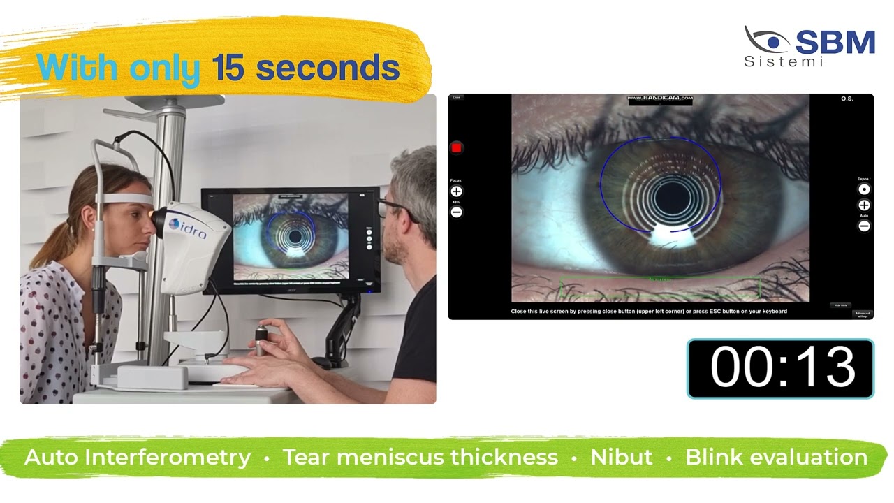
Thanks to one single video, the physician can gain lots of information: Automatic NIBUT; Average of more than one value; Graph to understand the trend of tear film stability during the video; Tear topography that shows all breaking the tear film during time.
Pupil diameter measurement is crucial in refractive surgery, as larger scotopic pupils may contribute to postoperative symptoms like halos, glare, and monocular diplopia. Accurate scotopic pupil measurements are also essential for determining optimal treatment zones in excimer laser, corneal, and intraocular surgeries.
Low tear production may result in aqueous tear deficiency (ATD) and cause dry eye symptoms. However, measuring the tear volume is difficult since the methods available nowadays are invasive and irritating.
MD. Luca Vigo of Studio Carones in Milan provides treatment suggestions based on their expertise in dry eye diagnosis and management. The IDRA system allows users to customize protocols and automatically suggest treatments after performing exams. The system includes a comprehensive database for tear film assessment, enabling ophthalmologists to diagnose deficiencies and determine appropriate treatments.
Evaluation of corneal diameter from limbus to limbus (white-to-white distance, WTW).
Images


Videos
Discover the perfect solution for you

basic
Interferomety test
Tear Meniscus
Auto NIBUT
-
Meibography
-
-
Blepharitis
Ocular redness classification
Pupillometry
White to White measurement
Anterior segment imaging
plus
Interferomety test
Auto Tear Meniscus
Auto NIBUT
NIBUT map and stability graph
Meibography
3D Meibography
Blink quality
Blepharitis
Ocular redness classification
Pupillometry
White to White measurement
Anterior segment imaging

full
Auto Interferomety test
Auto Tear Meniscus
Auto NIBUT
NIBUT map and stability graph
Meibography
3D Meibography
Blink quality
Blepharitis
Ocular redness classification
Pupillometry
White to White measurement
Anterior segment imaging


